Musculoskeletal

Perceptive is a leading provider of proven medical imaging services with extensive experience in musculoskeletal (MSK) imaging endpoint assessments. When tackling the many challenges involved in MSK trial imaging, global clinical trial sponsors repeatedly turn to Perceptive for the right experience, people, processes, and technology to drive their MSK development program’s success.

Two scientists discussing an image of a central nervous system scan on a computer screen.
divider

Perceptive MSK Trial Experience

Having supported over 360 MSK trials to date, many of which have led to regulatory approvals, Perceptive Imaging is uniquely positioned to deliver reliable imaging data to demonstrate the safety and efficacy of your compounds in development.

Expertise You Can Rely on

By working with Perceptive, you have access to experienced scientists and medical professionals who are experts in MSK imaging endpoints and criteria and are up to date on current, global regulations. They deliver technical expertise in the imaging modalities most often used in MSK trials, in protocol design, and in standardizing image acquisition across global investigative sites.

Arthritis / Rheumatology / Immunology

Spondyloarthritis

Radiographic Axial Spondyloarthritis (AS) is specifically characterized by the presence of structural damage in the sacroiliac joints (SIJs) which are noticeable on X-rays. This damage typically becomes visible several years after the initial onset of symptoms.

In contrast, non-radiographic axial spondyloarthritis (nr-AxSpA) does not display conclusive evidence of damage in the sacroiliac joints when examined through plain x-rays, unlike ankylosing spondylitis (AS). Instead, the presence of damage can only be visualized using MRI.

Perceptive brings extensive expertise in managing clinical trials within this field, having supported over 20 Ankylosing Spondylitis trials to date. Our team collaborates with renowned global experts who serve as central readers for these trials, ensuring the highest level of precision and accuracy.

Some of the criteria/assessments we have supported for Axial Spondyloarthritis trials are:

  • Modified New York Score (mNY)
  • ASAS OMERACT MRI Score
  • SPARCC for Spine and SIJ
  • Berlin Score
  • FASSS Scoring

Rheumatoid Arthritis (RA) and Psoriatic Arthritis (PsA)

X-ray and MRI-based assessments are used to support eligibility / stratification and / or efficacy endpoints.

X-ray is sometimes used for eligibility by confirming presence of bone erosions or juxta-articular bone formation (in support of deriving a CASPAR score for PsA). X-rays of joints in the hands and feet are used to derive scores for various criteria such as modified Van der Heijde Sharp (mTSS)  scores and other variations as needed to support efficacy endpoints.

MRI imaging is used to support Rheumatoid Arthritis MRI scoring (RAMRIS), Psoriatic Arthritis MRI scoring (PsAMRIS) and also in quantification of Rheumatoid Arthritis using MRI (RAMRIQ). In order to ensure consistency in positioning and image quality specialized hand positioners and templates are used. Additionally, MRI and ultrasound imaging (US) are employed to support inflammation in the joints such as in tenosynovitis etc.

Perceptive has supported the medical imaging needs of over 60 RA and PsA clinical trials to date, including the use of the following criteria/assessments:

  • Sharp Score
  • Van der Heijde Modified Sharp Score
  • Genant Modified Sharp Score
  • RAMRIS and PsAMRIS
  • RAMRIQ (Quantitative RAMRIS)

Osteoarthritis

Imaging in osteoarthritis trials is of critical importance as the endpoints relate to a reduction in joint space narrowing at the frequently evaluated joints (knee, hip, shoulder, and hands) as well as changes in cartilage and bone parameters. In order to enroll the right subjects several assessments are done both for inclusion and exclusion based on the trial design. This requires technical and imaging expertise as well as clear image acquisition guidelines and training of sites to acquire consistent images for X-ray and MRI.

Perceptive has supported the medical imaging needs of over 35 osteoarthritis trials and understands the specific needs of these trials.  For example, we provide special knee, hip, and hand positioners to sites and train the technicians to acquire the appropriate views and angles especially since Joint Space Width (JSW) is highly sensitive to the technique employed. MRI imaging in OA also requires dedicated protocol implementation on scanners and working closely with sites to provide timely feedback to optimize image quality.

Some of the criteria/assessments we have supported for Osteoarthritis trials are:

  • KL Grading
  • Joint Space Width
  • OARSI Scores
  • MOAKS / WORMS / BLOKS Scoring
  • Quantitative Cartilage and Bone Shape Assessments (through partnerships with expert third party vendors)

Women’s Health / Endocrine

Women’s Health

Perceptive has successfully supported a broad area of women’s health in indications such as endometriosis and uterine fibroids assessing efficacy and safety signals using ultrasound and DXA. DXA has been used to determine eligibility and safety such as bone loss and to support results reporting to sites /sponsors to facilitate treatment decisions during the trials. We have ultrasound and DXA expertise and oversight in authoring standardized guidelines as well as facilitating site training to ensure appropriate imaging and consistency.

Osteoporosis

Perceptive has supported 70 osteoporosis/bone mineral density trials with various drug candidates and biosimilars and has extensive scientific and logistical experience to successfully support such trials.

Dual-energy X-ray Absorptiometry (DXA) and X-ray imaging are routinely supported to assess efficacy and safety endpoints as well as support eligibility criteria. Our staff has extensive experience with Osteoporosis research/imaging, and we consult closely with clients to provide feedback on DXA and X-ray-related endpoints and safety signals during protocol development or revisions to ensure these are adequately captured. We also have a network of KOLs we can leverage to advise clients as needed.

DXA-derived T-scores as well as vertebral fracture assessments are reported to sites to enable timely decisions. Intelligent eCRF design and automated results reporting as per the unique requirements for each trial are put in place to ensure efficiency. In addition, our team also provides support to address site-central discordance and works closely with sponsors and sites to resolve such issues. Our solutions deployment and scientific staff work closely with the sponsor teams both to ensure the appropriate parameters and readouts are captured in the data exports and appropriate exports are designed to support any regulatory submissions.

Some of the criteria/assessments we have supported for Osteoporosis trials are:

  • Bone Mineral Density, T and Z Score Assessment by DXA
  • TBS Scores and Bone Quality
  • QCT Based Finite Element Analysis

Bone Age assessments in Growth Hormone deficiencies

Bone age serves as a means of interpreting skeletal maturity, holding significant importance in the comprehensive assessment and guidance of individuals with short or tall stature, impaired or accelerated growth, and delayed or early puberty. This evaluation is particularly relevant in pediatric studies focusing on issues such as obesity or growth hormone deficiencies.

An X-ray of the hand and wrist are acquired and assessed by comparing it to the Greulich and Pyle Atlas. At Perceptive, we have extensive expertise in this field and a knowledgeable team of readers. Additionally, we have the advantage of utilizing automated bone age assessment software (BoneXpert) based on the requirements of the trial.

Some of the criteria/assessments we have supported for Bone Age trials are:

  • Greulich and Pyle Bone Age Assessment (reader and / or automated software based)
  • Tanner Whitehouse and Fels using Bone Age Software (automated software based)

Metabolic / Body Composition Disorders

Gout

Perceptive experience with Gout imaging has included photography, X-ray, MRI, US and Dual Energy CT (DECT). DECT is used to detect the presence and quantify the extent of monosodium urate (MSU) crystals, which is a characteristic feature of tophaceous gout.

Our team is well-versed in the field of Gout, and we have the privilege of collaborating with Gout Key Opinion Leaders (KOLs) including Massachusetts General Hospital (MGH), who are renowned for their expertise in DECT analysis for Gout. Both our in-house experts and the KOLs are available for additional guidance to our clients as necessary.

Changes in Muscle / Fat / Body composition in various conditions

Perceptive has extensive experience with disorders related to muscle and fat changes such as Diabetes, Sarcopenia, Duchene Muscular Dystrophy, metabolic dysfunction, and Obesity-related changes in body composition measures related but not restricted to muscle and fat changes either in the whole body or at specific anatomic locations. Typically, most of these assessments are quantitative (volume or mass) with some being semi-quantitative.

We have worked with various modalities including DXA, CT, and MRI to delineate changes in these various anatomical locations. We can work on intra-muscular fat assessments using MRI sequences as appropriate and partner with third-party vendors on a need basis to facilitate the successful implementation of the right protocols and derive endpoints. We have dedicated expertise in-house to tailor sequences to achieve the desired outcomes in a standardized fashion which included direct communication with sites during initial protocol setup and robust QC to provide feedback.

Rare Diseases

Perceptive has experience in supporting various pediatric and adult rare indications affecting the musculoskeletal system such as Gaucher’s disease, Osteogenesis Imperfecta, Pompe disease, Duchenne Muscular Dystrophy, Spinal Muscular Atrophy (SMA), Achondroplasia and Fibrodysplasia Ossificans Progressive (FOP). We work closely with the client and expert readers/ KOLs to define the endpoints and design appropriate eCRFs to capture efficacy and safety endpoints to support decisions employing various modalities including but not restricted to X-ray, US, MRI, DXA, CT etc.

We tailor the image acquisition guidelines to ensure adequate coverage and appropriate views and implementation of scanning protocols. Toward this end, our experienced technical staff work closely with the sites to review protocol implementation, phantom scan acquisitions as appropriate, and tailored training. Given the nature of rare diseases and the unique image assessments involved, Perceptive is well positioned to provide guidance as well as work closely with relevant experts from the field as needed.

Some of the criteria/assessments we have supported for Rare Disease trials are:

  • Quantification of Heterotrophic Bone Ossification
  • MRI-based bone marrow burden Score (BMB score)
  • X-ray and MRI-based Assessments in Pediatric and Adult Populations for Rare Diseases
  • DXA based BMD Assessments
  • Variety of Qualitative Assessments based on Trial Endpoints
  • Muscle Quality and Volume

Meet our MSK Experts

By working with Perceptive, you have access to some of the industry’s most experienced MSK imaging experts. Our in-house medical imaging experts, including MDs, PhDs, imaging technologists, imaging physicists, and a dedicated exploratory imaging group are equipped and well-positioned to help you gain the information needed from new and novel imaging methods.

Sally (Sarah) Warner, PhD, CCD

Vice President, Scientific and Medical Services

Farhan Syed, PhD

Director, Scientific & Medical Services

Jener Basilio

Scientist

Perceptive's KOL Network

We collaborate with you to identify the most reliable readers and optimal read model for your trial, balancing their read volumes, and ensuring low inter-reader variability to drive the highest quality imaging reads for your study.

Our flexibility in choosing MSK-experienced readers enables you to utilize virtually any KOL as an expert reader including:

  • Prof. Jürgen Braun (Germany)
  • Dr. Kay-Geert Hermann (Germany)
  • Prof. Robert Landewe (Netherlands)
  • Prof. Désirée van der Heijde (Netherlands)
  • Dr. Charles Peterfy (USA)
  • Dr. William Palmer (USA)
  • Expert team at Massachusetts General Hospital


MSK Clinical Trial Modalities

Perceptive Medical Imaging delivers expertise in the significant majority of imaging modalities used to support the development of MSK-related treatments, including:

X-ray

X-ray imaging is a very common modality in a number of indications to support eligibility, efficacy, and safety such as in Ra/PsA, OA, OP etc.

Perceptive is well versed with the requirements and techniques / views needed for such assessments. Perceptive authors succinct and site/technologist friendly guidelines to allow for consistent and robust imaging which outlines the views and technical parameters such as KvP, collimation as well as the use of templates for consistent positioning etc.

Magnetic Resonance Imaging

MRI is a very versatile technique employed in a variety of indications involving the bones, muscles, and joints to assess structural and inflammatory changes at early and late stages of a disease or to monitor disease progression or resolution. Perceptive has successfully supported numerous studies using MRI read outs for various indications such as Ra, PsA, OA, AxSpA in support of eligibility as well as safety/efficacy reads.

Perceptive work closely with sites to implement the most appropriate protocols on MRI scanners at the sites as well as clearly outline requirements in the Image Acquisition Guidelines (IAGs). Our in-house MRI experts provide training and feedback to sites based on either phantom imaging or first subject scan quality to ensure appropriate scanning and quality.

CT / QCT

CT as a modality is used in a number of indications requiring quantitative or qualitative assessments of bone, fat and musculature at various levels in the anatomy. Calyx has done numerous trials looking at bony changes and quantification of osteophytes and heterotopic bone formation in indications such as Ankylosing Spondylitis as well as FOP. We also have supported single and multi-slice CT scans to determine muscle and fat content as well as to separate subcutaneous and visceral fat in clinical studies looking at metabolic changes and disorders such as in obesity, DMD, sarcopenia, Cachexia etc. Our trained staff thoroughly QC and ensure appropriate segmentation to facilitate quick and efficient central review by radiologists.

Perceptive also works closely with 3rd party vendors and KOL collaborators to derive quantitative CT measures in various skeletal abnormalities including assessment of volumetric BMD and estimation of bone strength and cortical and trabecular microarchitecture. We partner with ON Diagnostics to provide finite element analysis especially relevant for bone anabolic therapeutic drug candidates. We also partner with KOLs and academic labs to help obtain specialized and niche assessments such as trabecular and cortical architecture such as trabecular thickness and umbers and cortical thinning and porosity which yield critical information on short or long-term drug safety effects.

DXA / TBS

Dual Energy X-ray Absorptiometry (DXA) is the imaging modality of choice for clinical trials with changes in bone mineral density and /or body composition changes in the lean and fat mass compartments as an outcome measure such as in osteoporosis and obesity for example. DXA is critical to ascertain treatment effects on bone mass either for efficacy or for safety depending on the indication. For example, changes in hip and spine BMD are critical efficacy endpoints for drug candidates for osteoporosis while bone loss as a safety endpoint is employed in a number of indications such as endometriosis and fibroids in women or in inflammatory conditions such as Chron’s or adult and juvenile arthritis.

Body composition changes such as in bone, lean, or fat mass are also monitored by DXA in indications such as obesity, diabetes, HIV, etc. Perceptive has extensive experience in supporting all phases of global and region-specific DXA trials and we work closely with the clients and partners /sites to ensure that the appropriate imaging guidelines are adhered to and critical steps are done in a timely fashion such as Instrument quality control. Perceptive also partners with a third party vendor to provide TBS scores for trials looking at indices of bone quality. Perceptive’s experienced operational and scientific team closely engages with the client from the start of the trial to identify appropriate scanners, determine adequate training for technologists at sites and provide timely feedback on image quality.

Ultrasound

The application of ultrasound in various indications has continued to expand and is often used as a modality for many MSK indications.  Some indications where ultrasound has been used include hemophilia and osteoarthritis. Due to the improvement in the resolution of ultrasound transducers, diagnostic ultrasound in smaller joints has become more common in clinical and research settings.  Ultrasound does not use radiation and is painless and is an effective way to view both the structure and function of joint anatomy.  Ultrasound equipment is accessible, portable, and provides real-time visualization and assessment.

Given the complexity and required precision of MSK ultrasound imaging, Perceptive strongly recommends that images be obtained only by sonographers and physicians with significant MSK scanning experience.

Photography

Photography can be a valuable tool in evaluating and tracking inflammation, redness, and tophi in gout.  Photographs of hands and feet are most common with additional photos of elbows, knees, and other affected joints. Photography based assessment of tophus burden reduction can be performed using RECIST 1.1-like criteria using Computer-Assisted Photographic Evaluation in Rheumatology (CAPER) methodology.

Global Support

Perceptive’s operational model enables our client delivery teams to anticipate your study needs and scale our services to deliver quick and reliable continuity of service with efficient setup of sites, dedicated teams for QC support, and reduced imaging queries to meet your trial timelines.

With deep operational oversight delivered through MSK trial-experienced project managers, Perceptive Medical Imaging delivers the flexibility and scalability trial sponsors need during all stages of MSK treatment development, helping them to overcome challenges and confidently tackle unexpected trial changes.

With local support in India, China, North America, and Europe, you can have confidence that the imaging components of your MSK trial will run smoothly and deliver high quality data you can rely on. Every time.

Learn about Perceptive’s extensive imaging experience and capabilities in:


Oncology

Central Nervous System

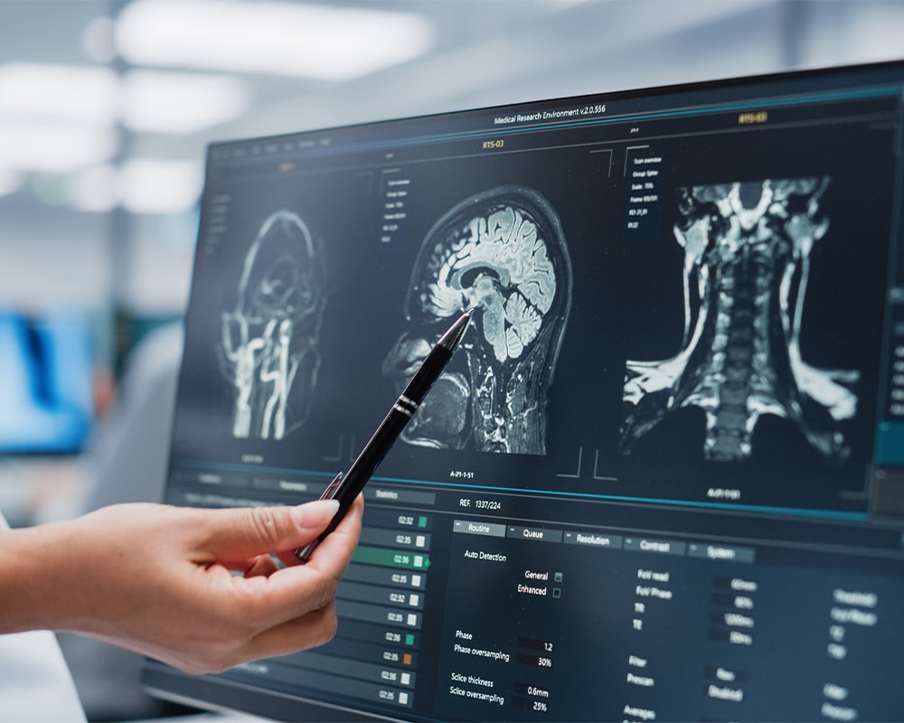
Person pointing at musculoskeletal image scan on computer screen with a ballpoint pen

Radiopharmaceuticals

Two detailed medical imaging scans of a human body, highlighting internal organs and systems with vibrant colours to represent various anatomical and functional regions, used for advanced diagnostic or dosimetry analysis.
divider